At Burwood eye clinic we partner with you in the management of your condition. Vision is so important to us, and the eye is a truly remarkable part of the body – we are learning more about it every day.
Your Consultation
Your Ophthalmic consultation will involve a detailed eye examination using specialised equipment. For a full examination we usually ‘dilate’ the pupil with eye drops to allow a thorough examination of the back of the eye.
We also conduct extra testing in the clinic to aid in the diagnosis and management of your condition.
The doctor will discuss the findings with you and together work out a management plan.
Investigations and Testing in the Clinic
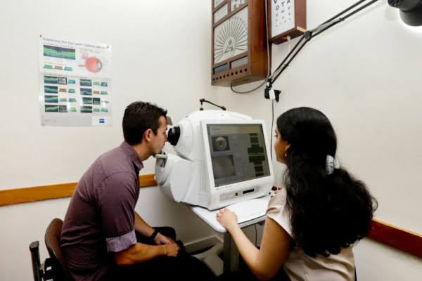
OCT stands for optical coherence tomography. This is a test we perform in the clinic which is quick, safe, non-invasive and reliable. It gives detailed pictures of the important structures at the back of the eye (in particular the macula, optic nerve and retinal nerve fibre layers) to help with the diagnosis and management of many important eye conditions.
At present there is no medicare rebate for these scans.
Computerised visual field testing is an important test of visual function. Many eye disorders, particularly glaucoma, and also other conditions, can affect the peripheral vision first. Often people may not be aware there are blind spots in the peripheral vision. This test helps to identify these, follow progress of the disease and response to treatment.
This test is performed in a darkened room. You will be asked to concentrate on a central light, and then click a button when you see other lights flashing off to the side. One of our experienced technicians will provide instructions, supervise and guide you as you perform the test. It gives valuable information in assessing for various eye diseases, and monitoring to see if the condition is controlled with treatment or deteriorating.
This is a scan of the front part of the eye which makes careful measurements of the curvature of the cornea. This helps diagnose various conditions at the front of the eye, and is also important in planning cataract surgery.
Prior to cataract surgery we conduct detailed scans to measure important parameters in the eye. This is to determine the power of the intraocular lens to be implanted when you have your cataract operation. Good preoperative measurements are one important ingredient in obtaining an optimal refractive outcome with cataract surgery. We first perform an IOLMaster scan which measures the curvatures at the front of the eye as well as the length of the eye. Sometimes we also use a special ultrasound to measure length in certain situations.

Laser Procedures in the Clinic
Selective laser trabeculoplasty is a treatment to lower the pressure for people with glaucoma and ocular hypertension. A drop is instilled to numb the eye, and a contact lens placed so that doctor can view the special part at the front of the eye where the aqueous fluid drains out. The procedure takes 5 mins and is relatively painless. It has a 70% chance of lowering the pressure, and is extremely safe. It is unusual for patients to have any problems after the laser, apart from a very transient mild discomfort. In some cases laser can be used instead of eye drops for treating glaucoma, in other cases it is used as well as drops – depending on stage of disease and severity.
In patients who have had cataract surgery sometimes a film can grow behind the artificial lens resulting in clouding of vision. The effect is sometimes a bit like the cataract ‘growing back’, but is a slightly different process. There is a relatively straight forward in office laser procedure to cut a hole in this skin and restore the vision once more. Roughly 50% of people who have had cataract surgery might require a laser capsulotomy at some point in future
This is where a small hole is made in the iris (coloured part of the eye surrounding the pupil) using a laser in the clinic.
This may be necessary in patients with angle closure glaucoma, or who are at risk of developing angle closure
This is a quick and relatively straightforward procedure. It is often done to prevent future problems in eyes that are at risk due to anatomical factors.
Surgical Procedures
Cataract surgery is one of the most commonly performed surgical procedures in the world. Modern cataract surgery is safe and effective. It is usually performed as a day procedure in a hospital, under local anaesthetic.
During surgery the cloudy lens is removed from the eye and replaced with an artificial intraocular lens. This also will change the focus of the eye so that people who require strong glasses may not need glasses for many tasks after the operation.
Cataract surgery can be life-changing and the vast majority of patients do very well. It is important to have a thorough examination before, however and to discuss in detail with your surgeon regarding the condition of your eyes and your particular circumstances, as well as the likely outcomes before proceeding.
Where drops or laser do not control the pressure enough glaucoma surgery may be required. While all Ophthalmologists treat glaucoma to some extent, cases where surgery is required is increasingly becoming a more specialised area of practice due to the complexities involved.
Trabeculectomy
This is an operation performed under local anaesthetic where a small hole is made at the top of the eye to allow fluid to drain out into the skin around the eyeball. When the fluid inside the eye drains into the ‘bleb’ that is formed under the eyelid the pressure will be at a lower and more consistent level. This is the closest we have to a cure for glaucoma – if successful patients do not need to use drops following the surgery. However in some people the body tries to heal over the hole that is made which will cause the pressure to go up again. Special anti-scarring medication is used at the time of surgery to help decrease the chance of trabeculectomy failure.
Just as important as the operation itself is the period of after care (at least 6-8 weeks, up to 3months) where relatively close outpatient follow up is required to ensure optimisation of the trabeculectomy bleb. Sometimes further interventions are required during this time. As with all surgeries there are risks, and some specific risks associated with trabeculectomy to be aware of before proceeding.
Tube Shunt Surgery
This is where a silicone tube is inserted into the eye to allow the fluid to drain and thus control the intraocular pressure. It may be recommended in eyes where previous surgery has not adequately controlled the pressure, or eyes with other conditions that make glaucoma surgery technically challenging. In eyes with more severe forms of glaucoma it is a very effective option, and relatively safe. Your doctor will discuss the risks and benefits prior to recommending any surgery.
Newer Devices for Glaucoma Surgery
There are an increasing number of new devices and techniques for glaucoma surgery, with a range of options now available for patients.
Dr. Wechsler has extensive experience in performing glaucoma surgery, and is also involved in trialling newer treatments as they become available.
We have been performing so called “minimally invasive” glaucoma surgical options such as the istent device, since their introduction in 2016.
A pterygium is a wing shaped piece of tissue that grows onto the cornea. The major risk factors are dry eye and sun exposure. These can cause irritation, be unsightly and in some cases can even interfere with the vision.
Pterygium surgery removes the abnormal tissue. This is done in a hospital usually under local anaesthetic as a day procedure. It is usually fairly straightforward to remove a pterygium however there is a risk of recurrence – where the pterygium grows back. To decrease the chance of recurrence a conjunctival autograft is used – this is where healthy conjunctiva from under the upper lid that has not been exposed to the sun as much is taken and secured over the bare area of sclera where the pterygium has been removed. This requires stitches or glue to be kept in place. Pterygium surgery can be painful in the days following surgery but we give strong pain killers to help cope with this until the eye heals. The stitches will be removed in the clinic at a postoperative visit later.
Many operations to treat corneal conditions require a form of transplantation. We are fortunate that the NSW eye bank co-ordinates the release of tissue from people who have kindly donated their eyes after they have passed away. There are strong protocols in place to ensure the supply of healthy corneal tissue for patients in need.
Because of the unique properties of the way the immune system interacts with the eye if you receive a corneal transplant you do not need to go on strong drugs to suppress the immune system to prevent rejection, as would happen for people who receive a kidney, heart or liver transplant, or other organs.
Endothelial Keratoplasty (DMEK, DSEK)
This is where a layer of endothelial cells taken from a donor cornea are placed inside the eye in place of the diseased layer of cells which is removed. This allows a swollen cornea to settle down and become clear again.
This is done through a small incision, and the recovery is usually fairly quick, almost like undergoing cataract surgery. It is done under local anaesthetic as a day procedure. Because it is only the internal layer that is replaced it does not weaken the eye structurally.
Deep Anterior Lamellar Keratoplasty (DALK)
In this procedure the top layers of the cornea are transplanted and the inner layer (containing the patients own endothelial cells) are left intact. There is less risk of rejection as compared to full thickness corneal transplant. This type of surgery is used to treat corneal problems that affect the front part of the cornea only, such as keratoconus.
In this operation stitches are used around the whole cornea to secure it in place. They have to stay in for at least 6 months, often more, to ensure the grafted cornea integrates into the patients eye.
Full Thickness Corneal Transplantation (PK – Penetrating Keratoplasty)
This is where the whole cornea is transplanted. This can treat a variety of corneal conditions. The graft is sutured in using multiple sutures which are left in until the graft has taken. It can take some time for maximial improvement in vision to occur following a corneal transplant, as there can be astigmatism induced form the corneal sutures. Also grafts can fail over time as there is a loss of endothelial cells in the process of surgery and postoperatively. Nevertheless corneal grafting can profoundly improve the outlook for patients with a variety of corneal conditions, and some corneal transplants performed by the surgeons at our clinic have been alive and well for over 30 years.
